Dr. Robert Ellsworth and Dr. Shawna Eischen highly recommend Thermography done at
Five Seasons Health by Pamela C. Mathews, CCT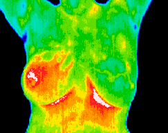
Call to schedule your Thermography appointment at (480)423-7060
A higher level of prevention specializing in breast imaging, pain diagnostics and early stage disease detection.
Thermal Imaging is painless, non invasive , has no compression and emits absolutely no radiation.
“With a multimodal approach, a woman’s chances for early breast cancer detection are 95%”
Please watch this very informative video regarding Medical Thermography and the benefits of early detection.
What it does: Breast thermography takes thermal images of the breasts and surrounding areas to aid in the early detection of breast cancer. Thermography assesses function rather than anatomically.
The principle: The procedure is based on the principle that chemical and blood vessel activity in both pre-cancerous tissue and the area surrounding a developing breast cancer is almost always higher than in the normal breast. Since pre-cancerous and cancerous masses are highly metabolic tissues, they need an abundant supply of nutrients (blood flow) to maintain their growth. In order to do this, they increase circulation to their cells by sending out chemicals to keep existing blood vessels open, recruit dormant vessels, and create new ones (neoangiogenesis). This process results in an increase in regional surface temperatures of the breast.
How it works: State-of-the-art breast thermography uses ultra-sensitive infrared cameras and sophisticated computers to detect, analyze, and produce high-resolution diagnostic images of these temperature and vascular changes.
Detection: The procedure is both comfortable and safe using no radiation or compression. By carefully examining changes in the temperature and blood vessels of the breasts, signs of possible cancer or pre-cancerous cell growth may be detected up to 10 years prior to being discovered using any other procedure. This provides for the earliest detection of cancer possible. Because of breast thermography’s extreme sensitivity, these temperature variations and vascular changes may be among the earliest signs of breast cancer and/or a pre-cancerous state of the breast.
Research: Breast thermography has been researched for over 30 years, and over 800 peer-reviewed breast thermography studies exist in the index-medicus.
In this database, well over 250,000 women have been included as study participants. The numbers of participants in many studies are very large– ranging from 37,000 to 118,000 women. Some of these studies have followed patients up to 12 years. Breast thermography has an average sensitivity and specificity of 90%.
Research Results: Studies show that:
* An abnormal infrared image is also the single most important marker of
high risk for developing breast cancer
* 8 times more significant than a first order family history of the disease.
* A persistently abnormal thermogram carries with it a 22x higher risk of future breast cancer.
* When added to a woman’s regular breast health checkups, a 61% increased survival rate has been realized.
* When used as part of a multimodal approach (clinical examination
+ mammography + thermography) 95% of early stage cancers will be detected.
Infrared Scan: A positive infrared scan may indicate the presence of many different breast abnormalities such as mastitis, benign tumors, fibrocystic breast disease, inflammatory breast disease, cancer and others.
Just as unique as a fingerprint, each patient has a particular infrared map of their breasts. Any modification of this infrared map on serial imaging (images taken over months to years) can constitute an early sign of an abnormality. In patients without cancer, the examination results are used to indicate the level of possible future cancer risk. Consequently, in the absence of other positive tests, an abnormal infrared image gives a woman an early warning. By maintaining close monitoring of her breast health with serial infrared imaging, self breast exams, clinical examinations, and other tests, a woman has a much better chance of detecting cancer at its earliest stage and preventing invasive tumor growth.
Unique Capability: Breast thermography’s ability to detect a pre-cancerous state of the breast, or signs of cancer at an extremely early stage, lies in its unique capability of monitoring the temperature variations and blood vessel alterations produced by the earliest changes in tissue physiology (function). At the earliest stages, a clinical thermographer can direct their client how to “clean up their inner environment” and change temperature variations by alkalizing, detoxing, changing their diet and teaching stress reduction techniques to reduce the inflammation.
Thermography vs other detections: Thermography assesses blood flow patterns, inflammation and function. Mammography, Ultrasound, Breast MRI assesses densities and anatomical masses. Thermography does not have the ability to visibly pinpoint the location of an early stage tumor. Mammography does not have the ability to see blood flow patterns and inflammation. Consequently, breast thermography is best used in conjunction to Mammography, Ultrasound, Breast MRI and/or physical examination when there is a concern. Since it has been determined that 1 in 8 women will get breast cancer, we must use every means possible to detect cancers when there is the greatest chance for survival. Proper use of breast self-exams, physician exams, thermography, and mammography together provide the earliest detection system available to date. If treated in the earliest stages, cure rates greater than 95% are possible.
Want to learn more about why traditional mammograms may not be the answer? Read this NBC news story to learn more!